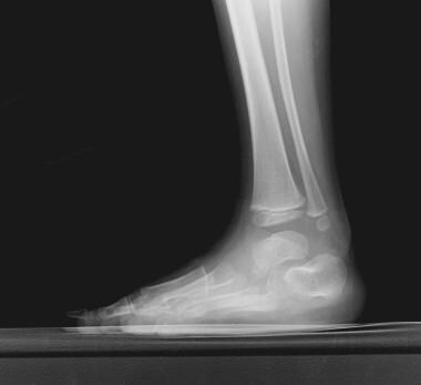
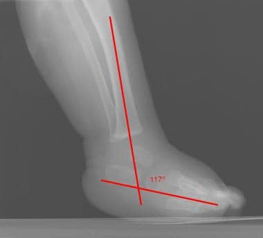
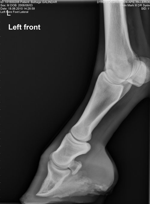
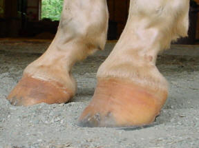
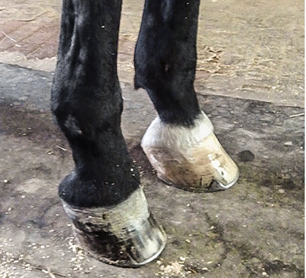
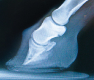
The radiograph below is from a weanling colt with a severe case of a "club foot" Figure 1 is the affected foot and figure 2 is the normal foot Xray vision was not necessary in this case to confirm the diagnosis due to the classic distortion of the hoof capsuleTerminology While some use talipes equinovarus and clubfoot synonymously, in certain publications, the term clubfoot is considered a more general descriptive term that describes three distinct abnormalities talipes equinovarus (adduction of the forefoot, inversion of the heel and plantar flexion of the forefoot and ankle);Radiograph video sequencing of a horse with displacement of the coffin bone due to laminitis (Andrew van Eps Chris Pollitt)

Equine Therapeutic Farriery Dr Stephen O Grady Veterinarians Farriers Books Articles
Club foot horse x ray
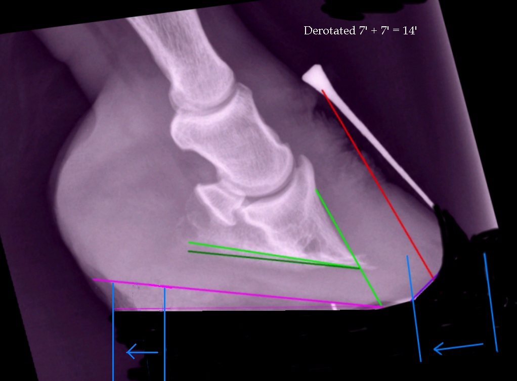
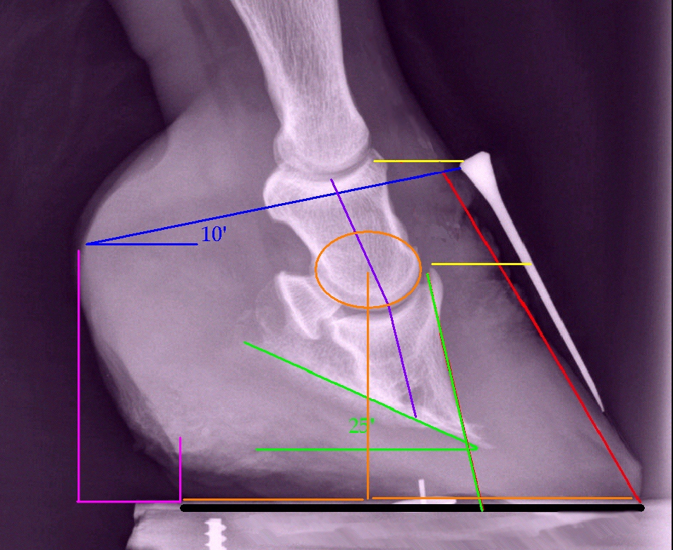
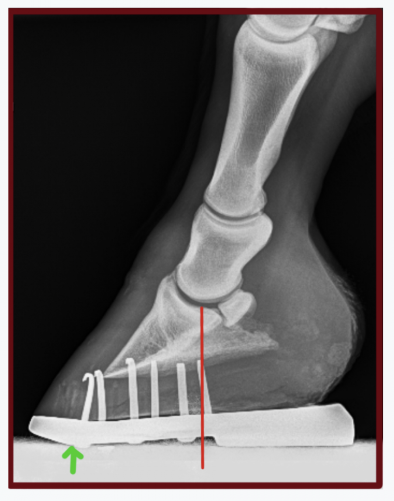
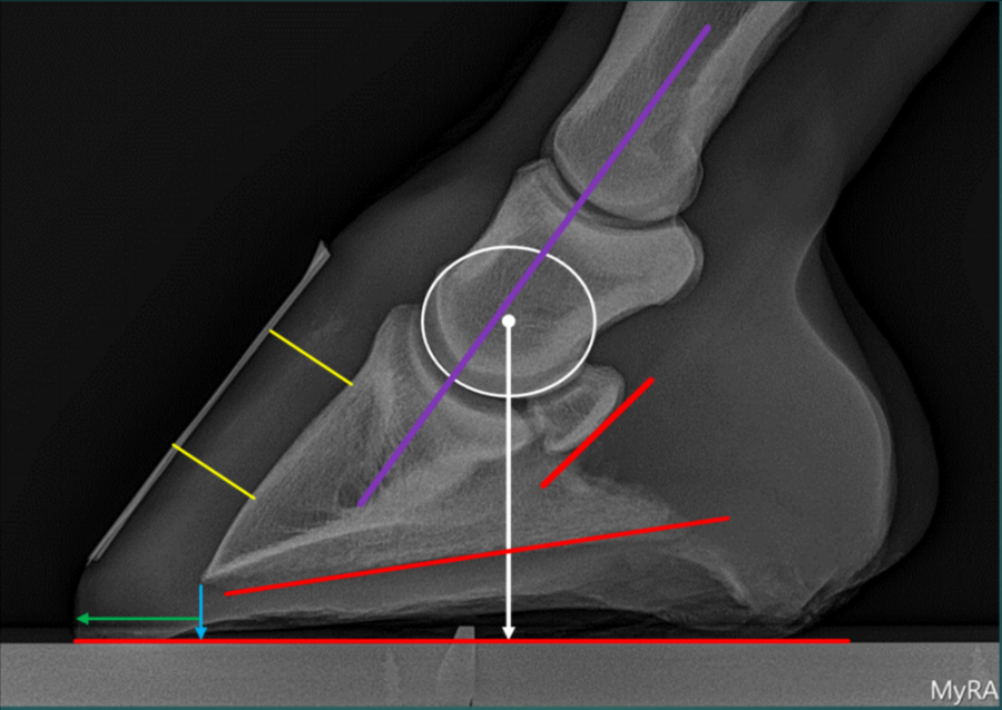
Club foot horse x ray-The search terms (horse* OR equine*) AND (foot OR feet OR digit* OR hoof OR hooves OR phalan * OR navicular) AND (radiograph* OR radiolog *) were generated and input into the PubMed search engine Following exclusion of studies more than 5 years old and those determined not to relate directly to the question, six useful results regardingThe xray will show whether the hoof pastern axis is parallel If the axis is broken forward (club foot) or if the axis is broken back (long toe underrun heel), the radiograph will reveal the degree of deformity and the best way to trim the foot to improve it Using landmarks, measurements can be drawn on the radiographs and transferred to the


Club And Subluxation In The Proximal Interfalangian Articulation Farriers Forum
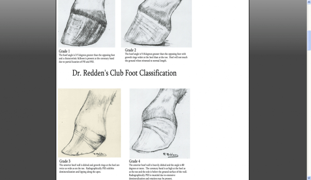
Seventyfour feet from 52 lame horses were included Twenty parameters were measured on radiographs, whereas the signal intensity, homogeneity and size of each structure in the foot were evaluated on magnetic resonance images The data were analysed using simple linear correlation analysis and classification and regression trees (CARTs) ResultsAn X ray of your horse's foot can help you predict the future while it shows you the present MECHANICAL LAMINITIS TREATMENT Foot X Rays A Crystal Ball?Correction of Club Foot (Inferior Check Ligament Desmotomy) A club foot is often characterized as a short toe and long heel combination A radiograph will reveal that P3, or the coffin bone, is rotated and not parallel with the ground Club feet have been classified into four grades 1 Slight rotation — May not be apparent to the eye 2
Club Foot Etiology congenital Imaging CAVE – Cavus (high arched foot), forefoot Adductus, hindfoot Varus / Equinus — AP radiograph overlap of talus and calcaneus, varus angulation of forefoot (metatarsus adductus), inversion of foot — Lateral radiograph parallel alignment of talus and calcaneus and equinus (upward) position of The difference is readily determined by physical and radiographic examination The normal pedal bone angle for most horses is between 42 and 48 degrees, when the physical angle of the pedal bones are greater than 48 degrees and both feet present with a more 'boxy' shape than normal, Club Foot Syndrome can be confidently diagnosedAny foot radiograph for alignment First, you can evaluate the relationship of the tibia to the hindfoot, then the relationship of the hindfoot to the midfoot, and finally the relationship of the midfoot to the forefoot As you read the case scenarios, you will see how this can be a
Any club foot that has been around a while will have a sensitive, unused, underdeveloped frog/digital cushion You can fix everything else and still have the back of the foot too sensitive for the horse to land on, which will cause the shortened stride and resulting club foot on its own – another vicious cycleBy Christy West, TheHorsecom Webmaster Article # 9805 When you look at a radiograph (X ray) of a horse's foot, do you visualize soft tissues, or do you only see bones? The contracted muscle/club foot condition is a common growth problem in young horses (up to 6 months of age), causing upright pasterns and a tiptoe stance This is often seen in foals with developmental problems due to rapid growth If discovered soon enough, this condition can be reversed by altering the foal's diet and reducing stress on


Clubfoot Imaging Practice Essentials Radiography Computed Tomography


Michael Porter Equine Veterinarian 12
Log In Log In Forgot Account?Labels club foot, club foot radiograph, equine podiatry, Sammy L Pittman DVM Sunday, Setting the bar for success in my laminitis cases Welcome to 14! To prepare the foot for radiographs, anesthetize the medial and lateral palmar digital nerves with mepivicaine 2% c at the level of the proximal sesamoids The horse should then be sedated with detomidine hydrochloride d (0002 001 mg/kg, IV) Clip and aseptically prepare the pastern in the area of the palmar digital vein


Low Foot Case Study Dixie S Farrier Service


Clubfoot Imaging Practice Essentials Radiography Computed Tomography
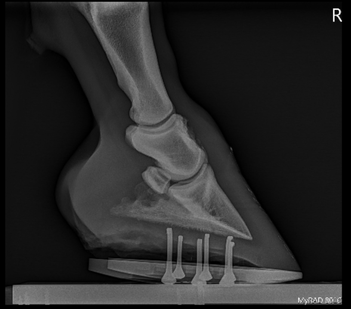
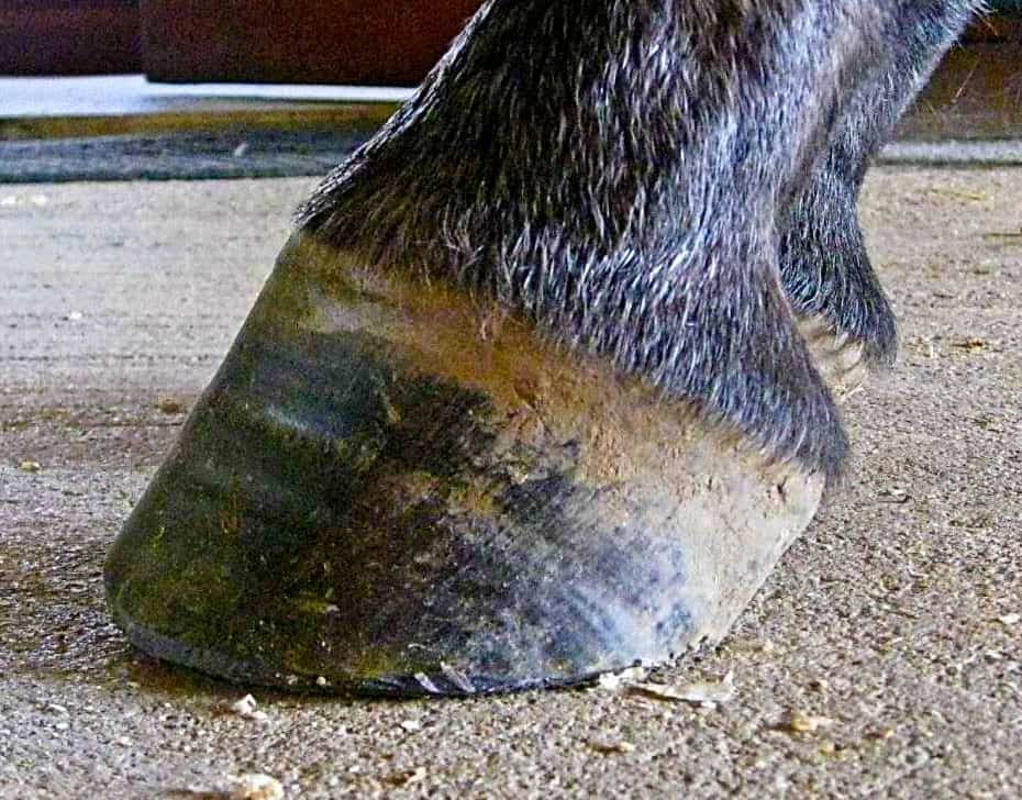
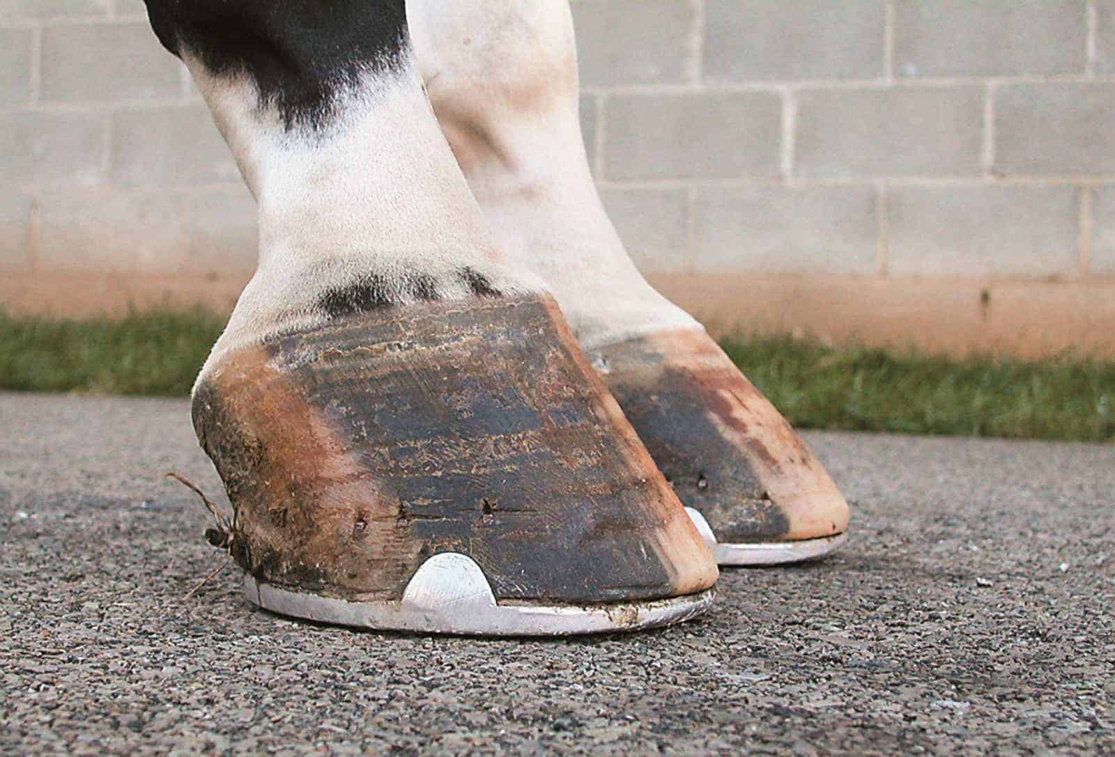
A horse with club foot has one hoof that grows more upright than the other The "up" foot is accompanied by a broken forward pastern, that is, the hoof is steeper than the pastern (Photo 1) In a normal foot, the hoof capsule and the pastern align Radiographs will show that the boneyOne area in which digital radiography has proved especially useful is working with farriers, especially on horses and ponies with laminitis We can now meet your farrier with the xray machine, and instantly show them the changes going on within your horse's foot This is invaluable in letting your farrier do the best job possibleRadiograph shows a moderate flexural deformity (yellow circle) involving the DIPJ in a horse with a club foot The flexural deformity is caused by a shortened DDF muscle tendon unit (red line) Grossly, the dorsal hoof wall angle is upright or steep accompanied by a broken forward foot


Recognizing And Managing The Club Foot In Horses Horse Journals


Understanding Club Foot The Horse Owner S Resource
A club foot generally a bit of a swear word in our house the frustration of the thought of a horse being present to me with a fault and always wondering if it could have been fixed earlier in life It's not that it's the end of the world for that horse It just up against it ,although over the last few months I have come across an ever Club Foot Conformation in Horses Caused by abnormal contraction of the deep digital flexor tendon, a club foot puts pressure on the coffin joint and initiates a change in a hoof's biomechanics Telltale signs of a club foot may include an excessively steep hoof angle, a distended coronary band, growth rings that are wider at the heels Dorsopalmar foot radiographs were acquired while varying the lateromedial stance;


Equine Podiatry Say What Mobile Veterinary Services


High Heels The Laminitis Site
If your horse is lame, your vet probably decided what area to radiograph based on the results of an examination, and blocks can be especially helpful When your vet blocks your horse, she'll inject a local anesthetic substance into nerves supplying an area, or directly into a joint or other enclosed structureAnd variable angle horizontal beam dorsopalmar foot radiographs were acquired while keeping the limb position constant Analyses of measurements demonstrated that hoof pastern angle had a linear relationship (R 2 = 0, P < 0Categories of club foot, on basis of joint motion and ability to reduce the deformities 11 i Soft foot also called postural foot can be treated by physiotherapy and standard casting treatment ii 2 Soft > Stiff foot occurs in 33% of cases It is usually a long foot which is more than 50% reducible and treated with


Hoof Evaluation Radiographs For The Farrier


Www Theneaep Com S Xia1nkm5hdp7yhwyucxfn39k42qa8m
Redden RF Radiographic imaging of the equine foot, Vet Clin North Am Equine Pract 03, 19 Weller R Radiography and radiology of the equine foot, in Proceedings 50th British Equine Veterinary Assn Congress 11, 50 29 Turner TA, Kneller SK, Badertscher RR, et al Radiographic changes in the navicular bone of normal horsesA club foot is a DEFORMITY and for any horse to win at top level competition it needs every possible advantage and no drawbacks The only way to stop continuing problems with club footed horses is not to breed from them After 11 months of gestation, it is a costly and heart breaking exercise if it results in a club footed foal Many articles have been written about club 'footed' horses Actually, horse do not have 'feet', dogs and humans do, but horses have hooves Therefore the term 'barefoot', as much as it is in common use now, really is a misnomer When we ride without hoof protection, we ride 'bare hoof' Ah well, a pet peeve


Podiatry Burwash Equine Services


Recognizing And Managing The Club Foot In Horses Horse Journals
In this edition of the IMV Imaging Journal Club, Clinical Manager Laura Quiney seeks to answer the question What is new in equine foot radiography?' A club foot alters a horse's hoof biomechanics, frequently leading to secondary lamenesses Affected horses tend to land toefirst, and their heel's growth rate isEquine Foot Management er) and Evaluating Radiographs for Equine Foot Management PDFs are extensively made use of as a way to existing information and facts with a hard and fast layout just like a paper publication Question Why generate a Evaluating Radiographs for Equine Foot Management PDF enewsletter?


Comstock Equine Hospital Veterinarian In City State Country Corrective Trimming


A Lateral And B Dorsopalmar Venograms Of A Normal Foot Areas Of Download Scientific Diagram
Symptoms of Club Foot in Horses Lameness Pain Excess toe wear Shortening of the tendon that is attached to the coffin bone Impacts the standing or movement of your young horse It can affect one or both limbs usually in the fore limbs Coronary band may bulge as the deformity progressesXray Scroll Stack Scroll Stack Lateral Lateral radiograph of the right foot shows that the long axes of the talus and calcaneus are nearly parallel The longitudinal arch is abnormally high AP radiograph of the right foot shows abnormally narrow talocalcaneal angle, with severe adduction and supination of the forefoot1/5 lame bilateral but 2/5 on left turn in a tight circle Left front is a grade 1 club and podiatry style films confirm healthy soft tissue parameters My thought process is With healthy sole depth and minimal remodelling of the apex of coffin bone on a club foot I want to next look at the navicular bone to evaluate for lesions


Contact Us Palmetto Equine Veterinary Services


Radiographic And Radiological Assessment Of Laminitis Sherlock 13 Equine Veterinary Education Wiley Online Library
Club Foot Horses Versus Uneven Weight Distribution First off let's discuss exactly what a "club foot" is This term is widely misused with regard to its use in horses with uneven hoof growth patterns The term "club foot" actually refers to a congenital defect of the foot and according to The Free Dictionary,Cuteness overload!😍How cute are they from 1 to 10?💞🎥 Unknown, DM me for credit!🍀Follow and horsesclubb for more!🍀#horse #horses #lovehorses #eqEquine Dental Specialty DENTAL RADIOGRAPHIC TECHNIQUES FOR THE HORSE This document is provided as an informational resource for AVDC Equine residents who are generating an equine dental radiograph set for Credentials Committee review It is not intended to be a comprehensive guide to equine dental and nasal radiography It is a companion


Severe Lameness Scarsdale Vets


High Heels The Laminitis Site
Talipes calcaneovalgus (dorsal flexion of the forefootI wanted to review some of my laminitis cases that have proven very successful with regards to quickly adding sole mass and demonstrating an even hoof wall growth from toe toThe horse should be positioned with both hind limbs fully weightbearing with the limb to be imaged slightly caudal to the contralateral limb (Figs 1–4) The xray beam should be directed horizontally, centered on the femorotibial joint, caudal and distal to the apex of the patella The beam will most com


Lesson 4


Talipes Equinovarus Clubfoot Radiology Case Radiopaedia Org
Radiograph shows a moderate flexural deformity involving the DIPJ in a horse with a club foot Note the ideal soft tissue parameters of the hoof capsule that can be assessed on this radiograph In older horses, when club foot has become a more chronic condition, altering the biomechanics of the foot is often the key to management Radiographs of the feet are essential in providing the information necessary to evaluate the existing biomechanics and determining the appropriate changes that need to be madeThe search terms (horse* OR equine*) AND (foot OR feet OR digit* OR hoof OR hooves OR phalan * OR navicular) AND (radiograph* OR radiolog *) were generated and input into the PubMed search engine Following exclusion of studies more than 5 years old and those determined not to relate directly to the question, six useful results were yielded


Equine Therapeutic Farriery Dr Stephen O Grady Veterinarians Farriers Books Articles


Hoof Conformation Vs Horse Conformation Scoot Boots Us Retail
A horse with an upright alignment of the pastern bones will also have upright hoovesa situation that is sometimes mistaken for club foot A true club foot is significantly more upright than the other hooves, or the angles of both hoof walls are steeper than the angles of the pasterns The severity of the problem is commonly graded on a fourThe equine club foot is a tricky issue Radiograph showing a flexion contracture of the coffin joint with increased hoof angle, often associated with dishing of the dorsal hoof wallInequality in the angles of a pair of hooves (usually front), ie one hoof grows steeper than the otherComes very useful in horses with upright feet, the best example being the clubfooted horse As the foot grows out in these horses, there is a propensity for the dorsal wall to distort and flare, producing multiple angles to the dorsal wall Radiographic evaluation of the dorsal wall with a conforming marker allows accurate assessment of the


Low Foot Case Study Dixie S Farrier Service


The Progressive Equine Services Hoof Care Centre Facebook
Clubfoot, or talipes equinovarus, is a congenital deformity consisting of hindfoot equinus, hindfoot varus, and forefoot varusThe deformity was described as early as the time of Hippocrates The term talipes is derived from a contraction of the Latin words for ankle, talus, and foot, pesThe term refers to the gait of severely affected patients, who walked on their anklesPedal Bone Fractures All You Need To Know Horse And Hound Pedal Bone Fracture In Horses Symptoms Causes Fracture Of Coffin Bone Generally Horse Side Vet Guide Parts Of The Horse Hoof The Coffin Bone Is The Predominant Bone Within The Equine Stabilizing Pedal Osteitis With A Glue On Shell AndSolution It is lowpriced


Club Foot Or Not Barefoot Hoofcare


Www Veterinariargentina Com Revista Wp284 Wp Content Uploads Radiographic And Radiological Assessment Of Laminitis Pages 524 E2 80 11 Pdf
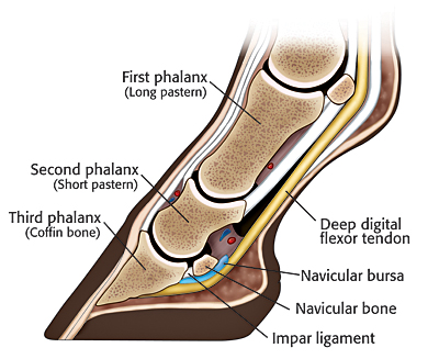
Radiographic examination of the foot is one of the most frequently performed studies of equine limbs 2 It is somewhat more complex than other radiographic studies of horse limbs, but with proper equipment and technique excellent quality radiographs can be made Without a good quality radiographic examination of the foot, correct radiographic diagnoses cannot be4 EQUINE VETERINARY JOURNAL Equine vet J (1984) 16 (l), 410 Com m issioned Articles Interpreting radiographs 2 The fetlock joint and pastern G B EDWARDS Department of Surgery and Obstetrics, Royal Veterinary College, Hawkshead House, Hawkshead Lane,Into consideration the horse's breed, age, environment, and use Preparing the foot Before taking any radiographs, the foot should be thoroughly cleaned of all dirt and debris, paying particular attention to the angle of the bars and to Fig 1 Lateral radiograph Notice both wings of P3 are superimposed (indicating good LM


Equine Podiatry Say What Mobile Veterinary Services


1
Clinical and Radiographic Examination of the Equine Foot 1 Introduction Lameness is one of the most frequently encountered problems in equine practice The foot


Recognizing And Managing The Club Foot In Horses Horse Journals


Laminitis Signalment Treatment And Prevention Flying Changes


ep Org Sites Default Files Issues Proceedings 12proceedings In Depth The Foot From Every Angle Eggleston Pdf


The Tolerable Club Foot The Horse


Boarding At Yucca Veterinary Medical Center


Managing The Club Hoof Easycare Hoof Boot News


Understanding X Rays The Laminitis Site


Q Tbn And9gct650ae6d8vnmqnvcvl4zcvlmqyfuvklxwislafu3ckq06siyee Usqp Cau


Surgical Services Scenic Rim Veterinary Service
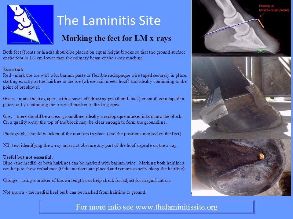


Understanding X Rays The Laminitis Site
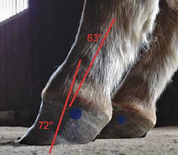


Defining And Fixing A Horse S Club Foot American Farriers Journal


Club And Subluxation In The Proximal Interfalangian Articulation Farriers Forum


ep Org Sites Default Files Issues Proceedings 12proceedings In Depth The Foot From Every Angle Eggleston Pdf


Hoof Evaluation Radiographs For The Farrier


Equine Podiatry In Wendell Nc Neuse River Equine Hospital


Rear Hoof Imbalance And The Effect On Rearlimb Lameness


The Wild Mustang Hoof
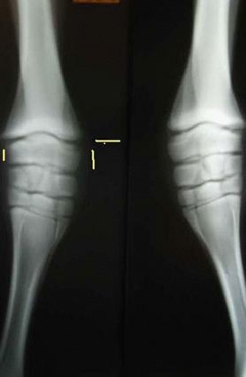


Types Of Crooked Legs In Foals
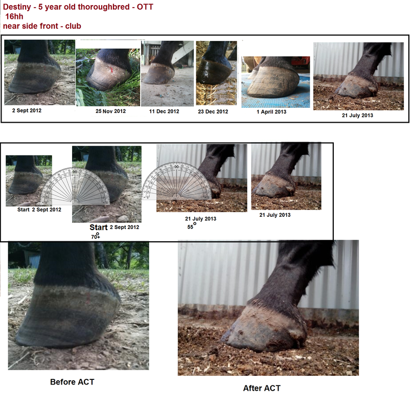


Club Foot Rehabilitation Act Trimming Strategy


Comstock Equine Hospital Veterinarian In City State Country Corrective Trimming


Farrier Terms Decoded Top 5 Farriery Terms Horse Owners Should Know Badger Veterinary Hospital


Cur Ost Normal And Not So Normal Horse Feet


Club Foot


The Chronicle Of The Horse


Equine Therapeutic Farriery Dr Stephen O Grady Veterinarians Farriers Books Articles


Equine Podiatry Dr Stephen O Grady Veterinarians Farriers Books Articles


Recognizing And Managing The Club Foot In Horses Horse Journals


Club Foot In Horses Brian S Burks Fox Run Equine Center Facebook


Radiolucency Glue On Horseshoes By Sound Horse Technologies


Shoeing Options For Club Foot In Horses


Club Foot Or Upright Foot It S All About The Angles American Farriers Journal


1


Managing The Club Foot The Horse


Understanding Navicular Syndrome Heel Pain In Horses


Www Theneaep Com S Xia1nkm5hdp7yhwyucxfn39k42qa8m


How To Treat Club Feet And Closely Related Deep Flexor Contraction


Natural Angle Volume 15 Issue 1 Spanish Lake Blacksmith


Recognizing Various Grades Of The Club Foot Syndrome


Recognizing And Managing The Club Foot In Horses Horse Journals


Equine Podiatry Dr Stephen O Grady Veterinarians Farriers Books Articles
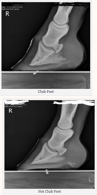


Managing The Club Hoof Easycare Hoof Boot News
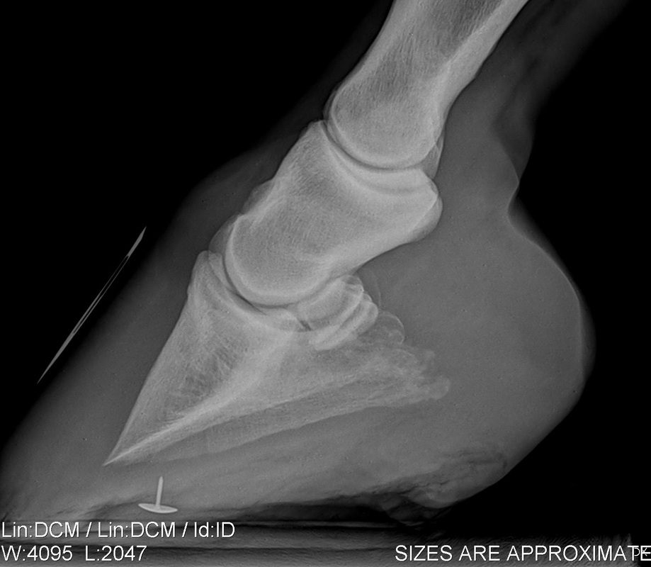


Understanding X Rays The Laminitis Site


Basic Shoeing Working With A Club Foot Farrier Product Distribution Blog


What S New In Equine Foot Radiology Imv Imaging


Shoeing Options For Club Foot In Horses


The Truth About Hoof Pastern Axis


Www Veterinariargentina Com Revista Wp284 Wp Content Uploads Radiographic And Radiological Assessment Of Laminitis Pages 524 E2 80 11 Pdf


Hoof Evaluation Radiographs For The Farrier


ep Org Sites Default Files Issues Proceedings 12proceedings In Depth The Foot From Every Angle Eggleston Pdf
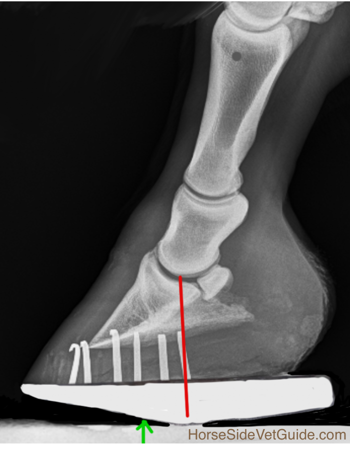


Understanding Navicular Syndrome Heel Pain In Horses


Clinical Radiology Of The Horse Medicine Health Science Books Amazon Com


ep Org Sites Default Files Issues Proceedings 12proceedings In Depth The Foot From Every Angle Eggleston Pdf


Farriers Archives Springhill Equine Veterinary Clinic


ep Org Sites Default Files Issues Proceedings 12proceedings In Depth The Foot From Every Angle Eggleston Pdf


No Foot No Horse Part 2 Hayes Equine Veterinary Services


Www Theneaep Com S Xia1nkm5hdp7yhwyucxfn39k42qa8m
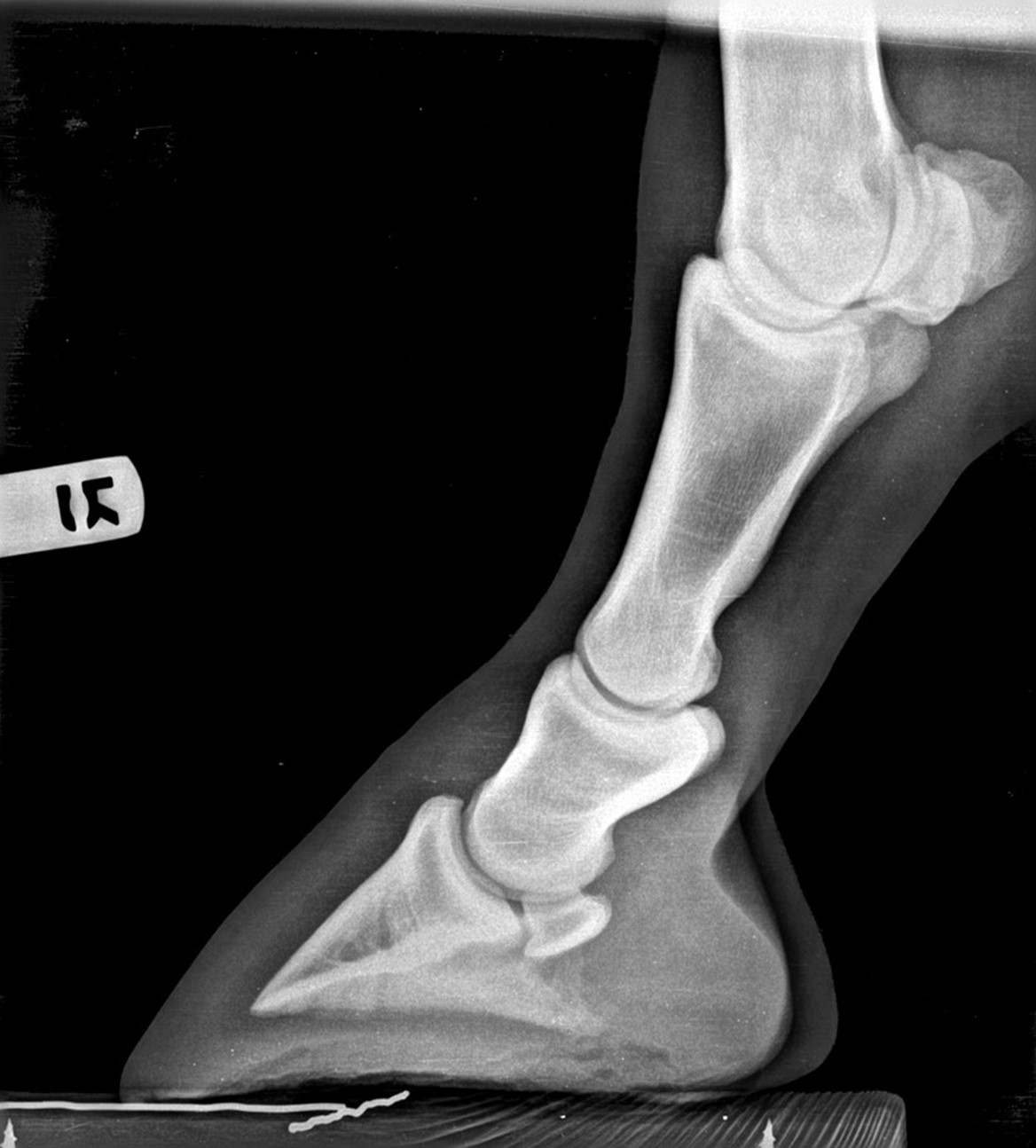


Hoof Radiographs Springhill Equine Veterinary Clinic


1


What Advice Has Been Most Helpful When You First Encounter A Club Foot American Farriers Journal


Epublishing


Recognizing And Managing The Club Foot In Horses Horse Journals


Www Theneaep Com S Dce80ecb B6af 4c6a 9a3f 0765cd Pdf


Pdf Value Of Quality Foot Radiographs And Their Impact On Practical Farriery Semantic Scholar


The Wild Mustang Hoof


Hoof Conformation Vs Horse Conformation Scoot Boots Us Retail


Thrush Cured Cowboy Pads And Copper Sulfate


Hoof Deformities Round Pen Square Horse


Radiographic Assessment Of The Ratio Of The Hoof Wall Distal Phalanx Distance To Palmar Length Of The Distal Phalanx In 415 Front Feet Of 279 Horses Mullard Equine Veterinary Education Wiley Online Library


Innovative Equine Podiatry Radiographic Parameters Measurement Guide


Performanceequinevs Com Wp Content Uploads 19 12 Equine Foot Secrets Ebook Compressed Pdf


Filly With Club Foot Barefoot Hoofcare


Laminitis A Pictorial Review


Podiatry Burwash Equine Services


Farriery For The Hoof With A High Heel Or Club Foot Semantic Scholar


Foal With Hoof Problems Club Foot The Horse


Basic Shoeing Working With A Club Foot Farrier Product Distribution Blog


0 件のコメント:
コメントを投稿